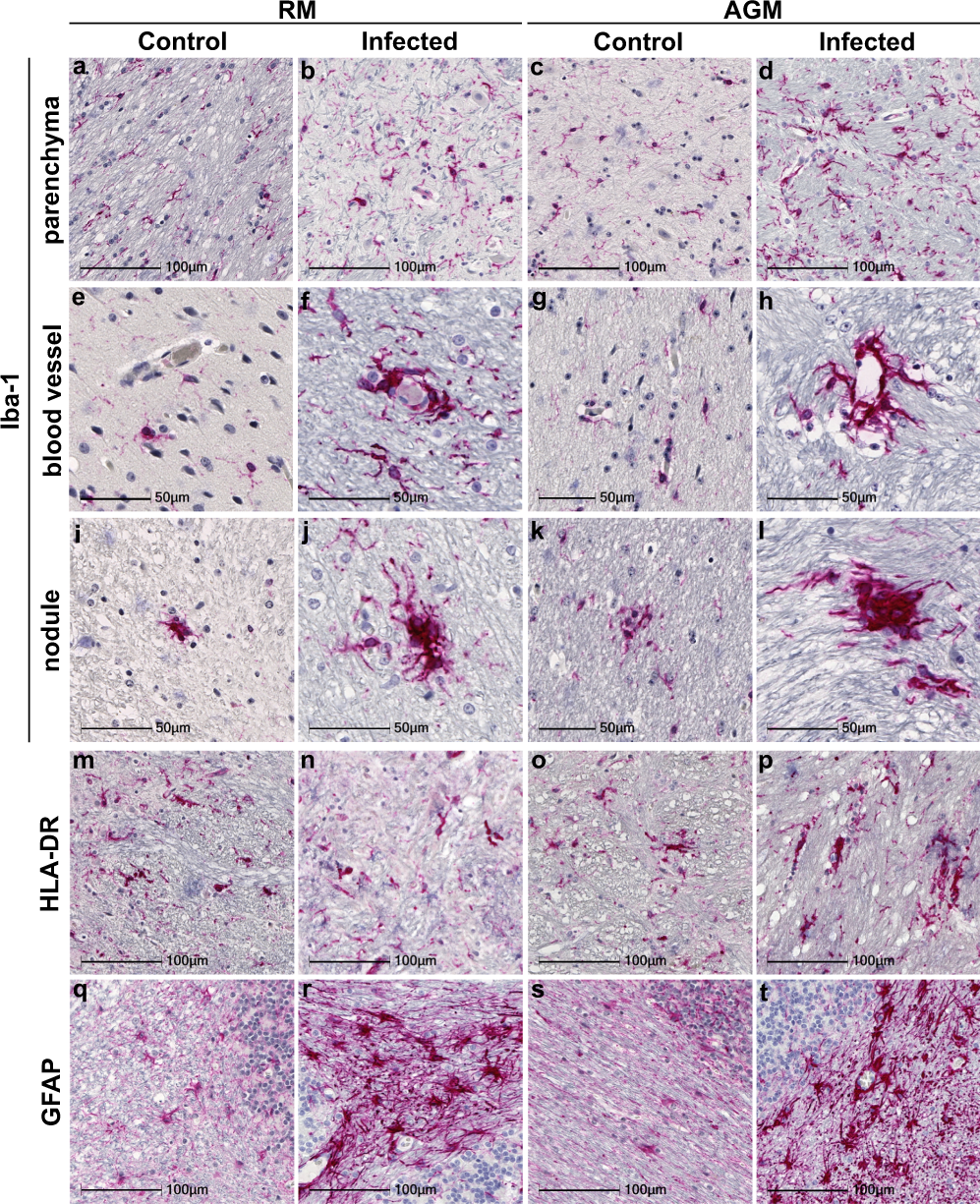
Fig. 1: Prominent neuroinflammation in brain of SARS-CoV-2 infected NHPs.
From: Neuropathology and virus in brain of SARS-CoV-2 infected non-human primates

Representative images identify microglia through Iba-1 immunopositivity in basal ganglia of mock-infected animals RM6 and AGM5 (a, c) that was upregulated in SARS-CoV-2 infected parenchyma, as shown in RM2 and AGM4 (b, d). Mild-moderate accumulation of microglia was often observed around blood vessels (RM1 f, AGM1 h). Nodular lesions were also frequently observed in brain of infected animals, represented here in RM4 and AGM4 (j, l). Microglial accumulation around blood vessels was not seen in age-matched mock-infected controls (RM6 e, AGM5 g), however, nodules (RM5 i, AGM5 k) were seen. These were less frequent and smaller than those observed in infection. Iba-1 immunopositivity also revealed morphological changes in microglia indicative of increased activation in infected animals, as compared to mock-infected controls, including large cell bodies with short, thickened processes (b, d, f, h, j, l). Microglial expression of HLA-DR was upregulated in the context of infection (n, p) seen in RM2 and AGM2, however, expression was also seen in control animals (m, o) represented by RM6 and AGM5. GFAP expression by astrocytes is upregulated and reveals morphological changes in the context of infection (cerebellum from RM4 r, AGM2 t), indicative of astrogliosis. Cerebellum from non-infected controls RM6 and AGM5 (q, s). Each immunohistochemical stain was performed twice on all brain regions. Abbreviations: AGM African green monkey, RM Rhesus macaque. Scale bars = 100 µm (a–d, m–t) and 50 µm (e–l).
