Abstract
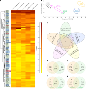
The impact of nanomaterial transformations on photosynthetic proteins remains largely unknown. We report positively charged iron oxide (Fe3O4) nanoparticles experience transformations in Arabidopsis thaliana plants in vivo that alter the formation and function of RuBisCO protein corona, a key carbon fixation enzyme. In vitro, negatively charged Fe3O4 nanoparticles impact the RuBisCO function but not their positively charged counterparts. Computational and in vitro proteomic analyses revealed that positively charged Fe3O4 nanoparticles preferentially bind to a RuBisCO small subunit that lacks active carboxylation sites. However, both positively and negatively charged nanoparticles decrease RuBisCO carboxylation activity after experiencing transformations in vivo by 3.0 and 1.7 times relative to the controls, respectively. The pH- and lipid-coating-dependent transformations that occur during nanoparticle transport across plant membranes enhance RuBisCO binding to positively charged nanoparticles affecting its distribution in chloroplasts. Elucidating the rules of how nanoparticle properties and transformations affect photosynthetic coronas is crucial for sustainable nano-enabled agriculture.
This is a preview of subscription content, access via your institution
Access options
Access Nature and 54 other Nature Portfolio journals
Get Nature+, our best-value online-access subscription
27,99 € / 30 days
cancel any time
Subscribe to this journal
Receive 12 print issues and online access
269,00 € per year
only 22,42 € per issue
Buy this article
- Purchase on SpringerLink
- Instant access to full article PDF
Prices may be subject to local taxes which are calculated during checkout


Data availability
Proteomics data for the proteomic analysis of NPs interfaced in vivo and in vitro are available at https://repository.jpostdb.org/entry/JPST003297.0 and for the partial digestion of RuBisCO with NPs are available at https://repository.jpostdb.org/entry/JPST003300. Any other data supporting the findings of this study are available in the Article and its Supplementary Information or from the corresponding author upon request. Source data are provided with this paper.
References
Cedervall, T. et al. Understanding the nanoparticle–protein corona using methods to quantify exchange rates and affinities of proteins for nanoparticles. Proc. Natl Acad. Sci. USA 104, 2050–2055 (2007).
Hadjidemetriou, M. & Kostarelos, K. Nanomedicine: evolution of the nanoparticle corona. Nat. Nanotechnol. 12, 288–290 (2017).
Mahmoudi, M., Landry, M. P., Moore, A. & Coreas, R. The protein corona from nanomedicine to environmental science. Nat. Rev. Mater. 8, 422–438 (2023).
Wheeler, K. E. et al. Environmental dimensions of the protein corona. Nat. Nanotechnol. 16, 617–629 (2021).
Borgatta, J. R. et al. Biomolecular corona formation on CuO nanoparticles in plant xylem fluid. Environ. Sci.: Nano 8, 1067–1080 (2021).
Pinals, R. L., Chio, L., Ledesma, F. & Landry, M. P. Engineering at the nano-bio interface: harnessing the protein corona towards nanoparticle design and function. Analyst 145, 5090–5112 (2020).
Prakash, S. & Deswal, R. Analysis of temporally evolved nanoparticle-protein corona highlighted the potential ability of gold nanoparticles to stably interact with proteins and influence the major biochemical pathways in Brassica juncea. Plant Physiol. Biochem. 146, 143–156 (2020).
Yu, Y., Dai, W. & Luan, Y. Bio- and eco-corona related to plants: understanding the formation and biological effects of plant protein coatings on nanoparticles. Environ. Pollut. 317, 120784 (2023).
Kurepa, J., Shull, T. E. & Smalle, J. A. Metabolomic analyses of the bio-corona formed on TiO2 nanoparticles incubated with plant leaf tissues. J. Nanobiotechnol. 18, 28 (2020).
Santana, I., Wu, H., Hu, P. & Giraldo, J. P. Targeted delivery of nanomaterials with chemical cargoes in plants enabled by a biorecognition motif. Nat. Commun. 11, 2045 (2020).
Spielman-Sun, E. et al. Protein coating composition targets nanoparticles to leaf stomata and trichomes. Nanoscale 12, 3630–3636 (2020).
Law, S. S. Y. et al. Polymer-coated carbon nanotube hybrids with functional peptides for gene delivery into plant mitochondria. Nat. Commun. 13, 2417 (2022).
Lowry, G. V. et al. Towards realizing nano-enabled precision delivery in plants. Nat. Nanotechnol. 19, 1255–1269 (2024).
Voke, E., Pinals, R. L., Goh, N. S. & Landry, M. P. In planta nanosensors: understanding biocorona formation for functional design. ACS Sens. 6, 2802–2814 (2021).
Bing, J., Xiao, X., McClements, D. J., Biao, Y. & Chongjiang, C. Protein corona formation around inorganic nanoparticles: food plant proteins-TiO2 nanoparticle interactions. Food Hydrocoll. 115, 106594 (2021).
Wong, M. H. et al. Lipid exchange envelope penetration (LEEP) of nanoparticles for plant engineering: a universal localization mechanism. Nano Lett. 16, 1161–1172 (2016).
Lew, T. T. S. et al. Rational design principles for the transport and subcellular distribution of nanomaterials into plant protoplasts. Small 14, e1802086 (2018).
Hu, P. et al. Nanoparticle charge and size control foliar delivery efficiency to plant cells and organelles. ACS Nano 14, 7970–7986 (2020).
Avellan, A. et al. Nanoparticle size and coating chemistry control foliar uptake pathways, translocation, and leaf-to-rhizosphere transport in wheat. ACS Nano 13, 5291–5305 (2019).
Zhang, Y. et al. Charge, aspect ratio, and plant species affect uptake efficiency and translocation of polymeric agrochemical nanocarriers. Environ. Sci. Technol. 57, 8269–8279 (2023).
Jeon, S.-J. et al. Targeted delivery of sucrose-coated nanocarriers with chemical cargoes to the plant vasculature enhances long-distance translocation. Small 20, e2304588 (2023).
Wu, H., Tito, N. & Giraldo, J. P. Anionic cerium oxide nanoparticles protect plant photosynthesis from abiotic stress by scavenging reactive oxygen species. ACS Nano 11, 11283–11297 (2017).
Husted, S. et al. What is missing to advance foliar fertilization using nanotechnology? Trends Plant Sci. 28, 90–105 (2023).
Jeon, S.-J. et al. Electrostatics control nanoparticle interactions with model and native cell walls of plants and algae. Environ. Sci. Technol. https://doi.org/10.1021/acs.est.3c05686 (2023).
Kim, K. et al. Sulfolipid density dictates the extent of carbon nanodot interaction with chloroplast membranes. Environ. Sci.: Nano 9, 2691–2703 (2022).
Zhu, L. et al. Cell wall pectin content refers to favored delivery of negatively charged carbon dots in leaf cells. ACS Nano 17, 23442–23454 (2023).
Dawson, K. A. & Yan, Y. Current understanding of biological identity at the nanoscale and future prospects. Nat. Nanotechnol. 16, 229–242 (2021).
Sharkey, T. D. The discovery of rubisco. J. Exp. Bot. 74, 510–519 (2023).
Giraldo, J. P. et al. Plant nanobionics approach to augment photosynthesis and biochemical sensing. Nat. Mater. 13, 400–408 (2014).
Swift, T. A. et al. Photosynthesis and crop productivity are enhanced by glucose-functionalised carbon dots. New Phytol. 229, 783–790 (2021).
Routier, C. et al. Chitosan-modified polyethyleneimine nanoparticles for enhancing the carboxylation reaction and plants’ CO2 uptake. ACS Nano 17, 3430–3441 (2023).
Bashiri, G. et al. Nanoparticle protein corona: from structure and function to therapeutic targeting. Lab Chip 23, 1432 (2023).
Sales, C. R. G., da Silva, A. B. & Carmo-Silva, E. Measuring Rubisco activity: challenges and opportunities of NADH-linked microtiter plate-based and 14C-based assays. J. Exp. Bot. 71, 5302–5312 (2020).
Xu, J. X., Alom, M. S., Yadav, R. & Fitzkee, N. C. Predicting protein function and orientation on a gold nanoparticle surface using a residue-based affinity scale. Nat. Commun. 13, 7313 (2022).
Wunder, T., Cheng, S. L. H., Lai, S.-K., Li, H.-Y. & Mueller-Cajar, O. The phase separation underlying the pyrenoid-based microalgal Rubisco supercharger. Nat. Commun. 9, 5076 (2018).
Poudel, S. et al. Biophysical analysis of the structural evolution of substrate specificity in RuBisCO. Proc. Natl Acad. Sci. USA 117, 30451–30457 (2020).
Payne, C. K. A protein corona primer for physical chemists. J. Chem. Phys. 151, 130901 (2019).
Li, G. et al. Association of heat-induced conformational change with activity loss of Rubisco. Biochem. Biophys. Res. Commun. 290, 1128–1132 (2002).
Kopac, T. Protein corona, understanding the nanoparticle-protein interactions and future perspectives: a critical review. Int. J. Biol. Macromol. 169, 290–301 (2021).
Botella, C., Jouhet, J. & Block, M. A. Importance of phosphatidylcholine on the chloroplast surface. Prog. Lipid Res. 65, 12–23 (2017).
Reszczyńska, E. & Hanaka, A. Lipids composition in plant membranes. Cell Biochem. Biophys. 78, 401–414 (2020).
Leibe, R. et al. Key role of choline head groups in large unilamellar phospholipid vesicles for the interaction with and rupture by silica nanoparticles. Small 19, e2207593 (2023).
Ganguly, S. & Margel, S. Bioimaging probes based on magneto-fluorescent nanoparticles. Pharmaceutics 15, 686 (2023).
Hoang, K. N. L., Wheeler, K. E. & Murphy, C. J. Isolation methods influence the protein corona composition on gold-coated iron oxide nanoparticles. Anal. Chem. 94, 4737–4746 (2022).
Pu, S., Gong, C. & Robertson, A. W. Liquid cell transmission electron microscopy and its applications. R. Soc. Open Sci. 7, 191204 (2020).
Schmidt, R. et al. MINFLUX nanometer-scale 3D imaging and microsecond-range tracking on a common fluorescence microscope. Nat. Commun. 12, 1478 (2021).
Gasteiger, E. et al. in The Proteomics Protocols Handbook 571–607 (Humana Press, 2005).
Sharkey, T. D., Bernacchi, C. J., Farquhar, G. D. & Singsaas, E. L. Fitting photosynthetic carbon dioxide response curves for C3 leaves. Plant Cell Environ. 30, 1035–1040 (2007).
Wang, Y. & Hernandez, R. Construction of multiscale dissipative particle dynamics (DPD) models from other coarse-grained models. ACS Omega 9, 17667–17680 (2024).
Deng, C. et al. Nanoscale iron (Fe3O4) surface charge controls Fusarium suppression and nutrient accumulation in tomato (Solanum lycopersicum L.). ACS Sustain. Chem. Eng. 12, 13285–13296 (2024).
Wu, M. et al. Solution NMR analysis of ligand environment in quaternary ammonium-terminated self-assembled monolayers on gold nanoparticles: the effect of surface curvature and ligand structure. J. Am. Chem. Soc. 141, 4316–4327 (2019).
Mahajan, S. & Tang, T. Martini coarse-grained model for polyethylenimine. J. Comput. Chem. 40, 607–618 (2019).
Acknowledgements
This work was supported by the National Science Foundation under grant no. CHE-2001611, the NSF Center for Sustainable Nanotechnology. We acknowledge support from the UC President’s Pre-Professoriate Fellowship to C.C. We thank J. Arrington from the University of Illinois Urbana-Champaign for processing the raw proteomic data, and M. M. Dickinson from the Central Facility for Advanced Microscopy and Microanalysis at the University of California, Riverside, for the TEM analysis sample preparation. We also thank the UC Riverside Metabolomics Core for performing the metabolomics analysis. The computing resources necessary for this work were performed in part on Bridges at the Pittsburgh Supercomputing Center through allocation CTS090079 provided by the Advanced Cyberinfrastructure Coordination Ecosystem: Services & Support (ACCESS), which is supported by the National Science Foundation (NSF) grant nos. 2138259, 2138286, 2138307, 2137603 and 2138296. Additional computing resources were provided by the Advanced Research Computing at Hopkins (ARCH) high-performance computing (HPC) facilities.
Author information
Authors and Affiliations
Contributions
J.P.G. and C.C. conceived the idea, designed the experiments and performed the data analysis. S.-J.J. conducted the fluorescence imaging analysis to visualize the in vivo corona formation on NPs and performed the biocompatibility measurements. C.J.M. and K.N.L.H. contributed to the proteomic analysis and performed the in vitro RuBisCO activity measurements. C.A. performed the pH-dependent in vitro RuBisCO protein corona formation experiments. K.E.W. and C.A. performed and analysed the samples for circular dichroism. K.E.W. and E.S. contributed with the proteomic analysis support. J.C.W., C.D. and Yi Wang collected and analysed the TEM images. R.H., X.W. and Yinhan Wang performed the coarse-grained simulations and analysis. All authors contributed to the manuscript writing.
Corresponding author
Ethics declarations
Competing interests
The authors declare no competing interests.
Peer review
Peer review information
Nature Nanotechnology thanks Renu Deswal, Yaning Luan and the other, anonymous, reviewer(s) for their contribution to the peer review of this work.
Additional information
Publisher’s note Springer Nature remains neutral with regard to jurisdictional claims in published maps and institutional affiliations.
Supplementary information
Supplementary Information
Supplementary Figs. 1–17, Methods, Results and References.
Supplementary Tables 1–19
Supplementary Tables 1–19.
Supplementary Source Data
Source data for supplementary figures.
Source data
Source Data Fig. 2
Source data for Fig. 2.
Source Data Fig. 3
Source data for Fig. 3.
Source Data Fig. 4
Source data for Fig. 4.
Source Data Fig. 5
Source data for Fig. 5.
Source Data Fig. 6
Source data for Fig. 6.
Source Data Fig. 5
Unprocessed SDS-PAGE gels for Fig. 5a,b.
Source Data Fig. 6
Unprocessed SDS-PAGE gels for Fig. 6a–d.
Rights and permissions
Springer Nature or its licensor (e.g. a society or other partner) holds exclusive rights to this article under a publishing agreement with the author(s) or other rightsholder(s); author self-archiving of the accepted manuscript version of this article is solely governed by the terms of such publishing agreement and applicable law.
About this article
Cite this article
Castillo, C., Jeon, SJ., Hoang, K.N.L. et al. In vivo transformations of positively charged nanoparticles alter the formation and function of RuBisCO photosynthetic protein corona. Nat. Nanotechnol. (2025). https://doi.org/10.1038/s41565-025-01944-x
Received:
Accepted:
Published:
DOI: https://doi.org/10.1038/s41565-025-01944-x


